
100 consecutive patients with papillary carcinoma - Case 21. (ultrasonographic picture 9)
|
|

|
|
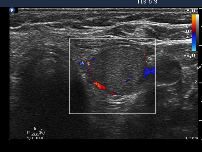
Lower part of the left lobe, horizontal scan, color Doppler mode. There are signs of a perinodular blood flow.