
100 consecutive patients with papillary carcinoma - Case 27. (ultrasonographic picture 5)

|

|

|
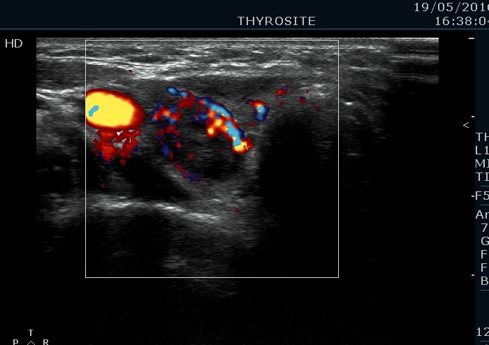
Lower part of the right lobe, horizontal view, color Doppler mode. This is a suspicious vascular pattern.


