
100 consecutive patients with papillary carcinoma - Case 33. (ultrasonographic picture 8)
|
|

|
|
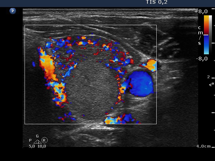
Left lobe, horizontal scan, color Doppler mode. This pattern pretends as to the nodule had perinodular blood flow, while in fact only the extranodular part of the lobe presents markedly increased vascularization. The nodule displays neither perinodular nor intranodular blood flow.