
100 consecutive patients with papillary carcinoma - Case 34. (ultrasonographic picture 5)
|
|

|
|
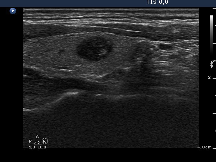
Left lobe, longitudinal scan. The acoustic shadow proves that the smaller lesion contains not only microcalcification but coarse calcification, as well. This nodule proved to be benign on histopathological analysis.