
100 consecutive patients with papillary carcinoma - Case 34. (ultrasonographic picture 7)
|
|

|
|
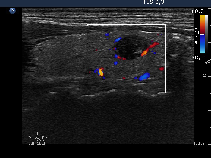
Left lobe, longitudinal scan, color Doppler mode. The nodule, that proved papillary carcinoma, presents signs of a perinodular blood flow.