
100 consecutive patients with papillary carcinoma - Case 39. (ultrasonographic picture 5)

|

|

|
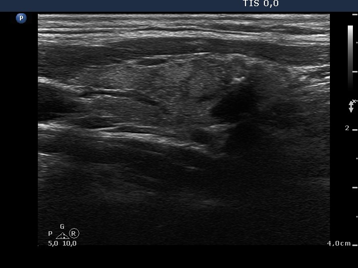
Left lobe, longitudinal scan. There is a lesion in the lower two-third of the lobe. The borders are irregular. Note the presence of non-specific granules and more bright microcalcifications.


