
100 consecutive patients with papillary carcinoma - Case 42. (ultrasonographic picture 1)

|

|
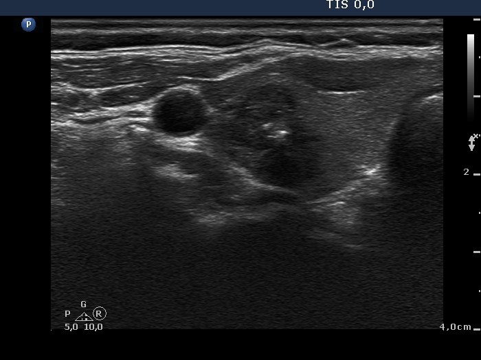
Right lobe, horizontal view. There is a hypoechogenic lesion composed of at least two nodules. The medial borders of the mass are partly blurred, partly lobulated. The interpretation of echogenic figures is equivocal, they might be either microcalcifications or presentation of connective tissue based on the synchronous presence of echogenic lines and granules.


