
100 consecutive patientswith papillary carcinoma - Case 47. (ultrasonographic picture 7)

|

|

|
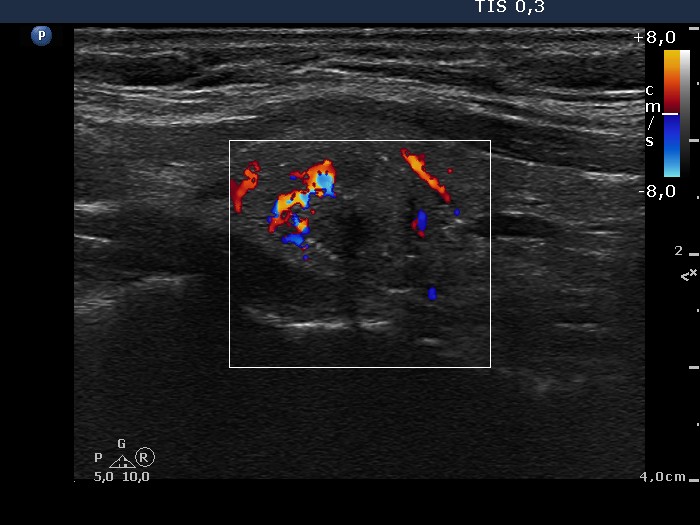
Lower part of the left lobe, longitudinal scan, color Doppler mode. The lesion presents an irregular vascular pattern.


