
100 consecutive patients with papillary carcinoma - Case 50. (ultrasonographic picture 5)

|

|

|
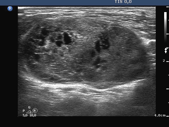
Lateral to the right lobe, horizontal scan. There is a large hypoechogenic mass presenting cystic degeneration. Note the difference in the echo structure between the lateral (left in the image) and the medial part (right in the image) of the mass. The latter is more hypoechogenic and contained the malignant focus.


