
|
|
100 consecutive patients with papillary carcinoma - Case 62.
|

|


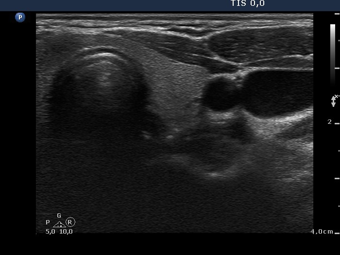


Clinical data: A 34-year-old woman was referred for evaluation of a thyroid nodule, which was discovered by the patient herself 3 months ago.
Palpation: a very firm nodule in the right side of the isthmus.
Functional state: euthyroidism (TSH 1.56 mIU/L).
Ultrasonography. The thyroid was echonormal. There was a moderately hypoechogenic nodule in the right side of the isthmus. The ventral part of the lesion was not separated from the sternocleidomastoid muscle. The lesion presented microcalcifications and signs of perilesional blood flow.
Cytology resulted in papillary carcinoma.
A right lobectomy was performed. Histopathology disclosed a T1N0 papillary carcinoma with a maximal diameter of 8 mm.
Comment. The ultrasound pattern was highly suspicious of extrathyroidal spread of the tumor.


