
|
|
Study on extrathyroidal spread of papillary carcinoma - Case 6.
|

|


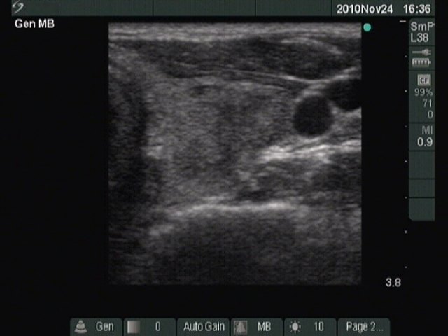
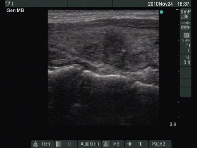
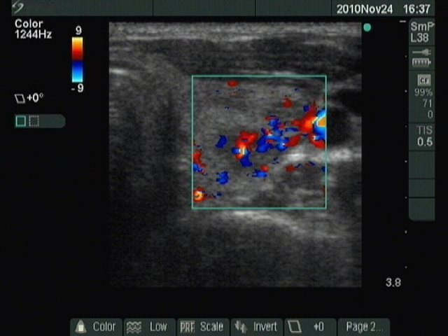
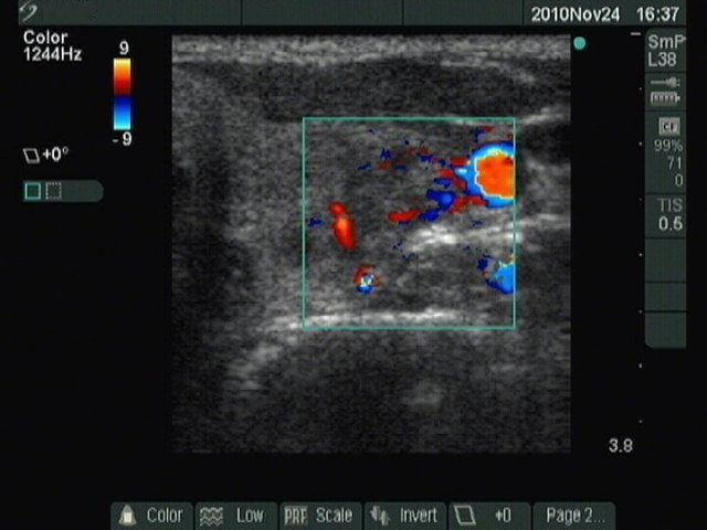
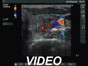


Initial examination (first row of images)
Clinical data: a 41-year-old woman referred for evaluation of weight gain.
Palpation: the left thyroid was tender, no nodule was palpable.
Functional state: euthyroidism with TSH 0.82 mIU/L, FT4 13,7 pM/L, anti-TPO 1.8 U/mL.
Ultrasonography. The thyroid was echonormal. There was a hypoechogenic area in the dorsolateral portion of the left lobe. The lesion had irregular, blurred borders, contained one focus of microcalcification and presented an irregularly increased intralesional flow.
Cytology of the nodule resulted in benign lesion with signs of inflammation not otherwise specified.
We advised a repeat examination in a year.
Second examination 4 years later (second row of images)
Clinical data: the patient had no complaints therefore she visited us only four year after the initial examination.
Palpation: the left thyroid was tender, no nodule was palpable.
Functional state: euthyroidism with TSH 1.08 mIU/L, anti-TPO 0.3 U/mL, CRP 0 mg/dL.
The lesion in question is located in the central part of the left lobe


