
|
|
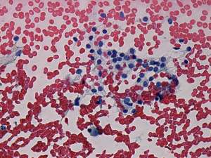
Study on extrathyroidal spread of papillary carcinoma - Case 11.
|
|


First examination (first row of sonographic images):
Clinical presentation: a 33-year-old woman was referred for an evaluation of typical complaints suggesting hyperthyroidism.
Palpation: both thyroids were enlarged.
Functional state: hyperthyroidism (TSH undetectable, FT4 42.2 pM/L, FT3 above 46 pM/L).
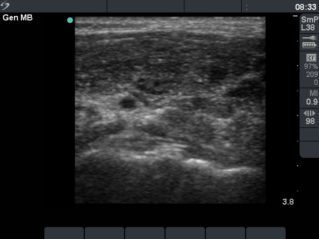
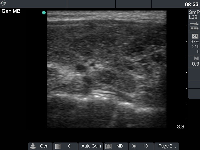
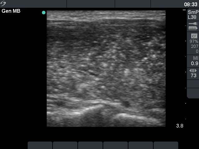
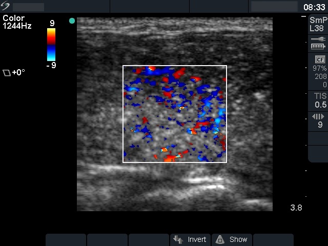
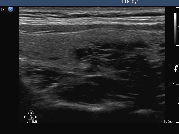
Ultrasonography: the thyroid was diffusely hypoechogenic. The vascularization was increased. The left thyroid was significantly more inhomogeneous than the right lobe. Compare the first two index pictures in the first row with the third and fourth one, right and left lobe, respectively.Thyrostatic drug was administered for 12 months, thereafter the drug was discontinued.
Follow-up examination 17 months after first visit (second and third rows):
Clinical presentation: the patient was well. She came to sonographic follow-up suggested at her first visit.
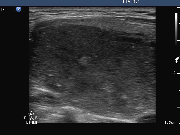
Palpation: a hard nodule in the upper part of the left thyroid.
Functional state: subclinical hyperthyroidism (TSH undetectable, FT4 21.1 pM/L, FT3 6.68 pM/L, TSAB 1.9 U/L (normal value below 1.5)).
The lesion in question was located in the upper 2/3 of the left lobe.