
|
|
100 consecutive patients with thyroid nodule - Case 1.
|
|


First examination (first and second rows of images)
Clinical presentation: a 56-year-old man was referred for follow-up examination of a thyroid nodule. The patient was known harboring a nodule for 4 years. Previous cytology from the "dominant nodule in the left thyroid" resulted in benign lesion in another institute. The patient had no complaints.
Palpation: a firm nodule in the right lobe and a nodular part in the left thyroid.
Functional state: euthyroidism (TSH 1.33 mIU/L).
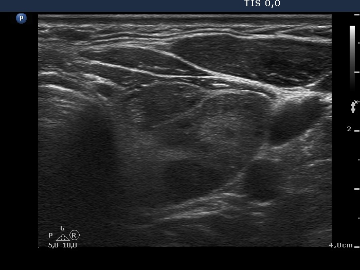
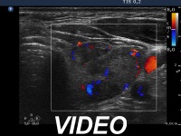
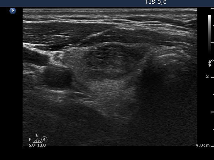
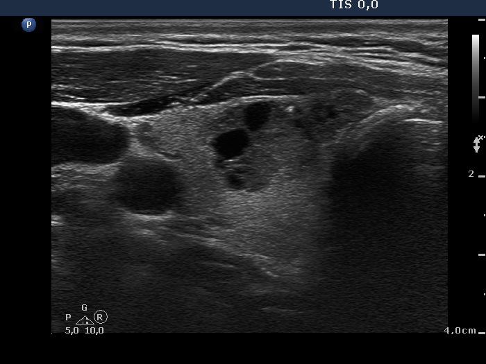
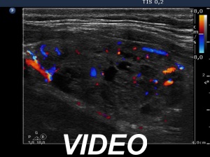
Ultrasonography. The thyroid was echonormal. There were two moderately hypoechogenic nodules in the ventromedial part of the right lobe. One of them presented irregular borders, hyperechogenic granules and cystic degeneration. The left thyroid contained several nodules with different echo structures.
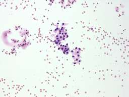
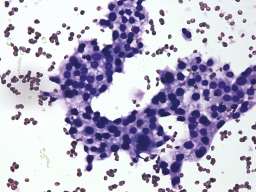
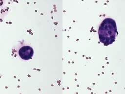
Cytology was performed from the nodule in the right lobe and resulted in benign, follicular proliferation..
Second examination 3 years later (third row of images)
Clinical presentation: the patient had no complaints.
Palpation: a moderately firm nodule in the right thyroid.
Functional state: euthyroidism (TSH 1.41 mIU/L).
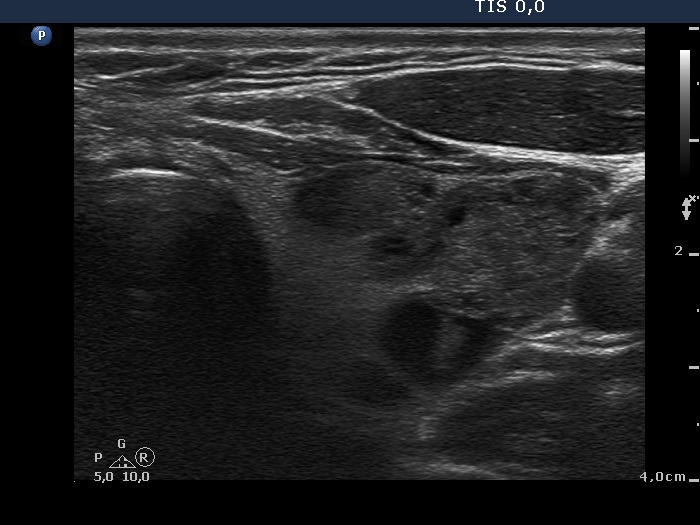
Ultrasonography. No significant changes could be found. The size of the nodule in the right lobe and that of the multinodular left lobe was unchanged.
Comment. This example emphasizes the importance of the measurement of the volume of the thyroid lobes. The left lobe contained multiple nodules next to each other. Neither of these lesions can be measured correctly. To basis of the follow-up is therefore the volume of the entire lobe and not that of one or another nodule.
.