
100 consecutive patients with thyroid nodule - Case 13. (ultrasonographic picture 7)

|

|

|
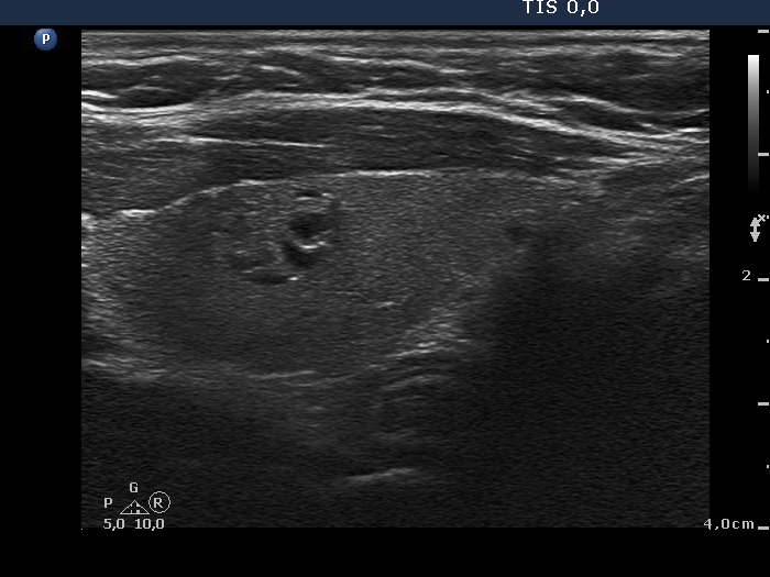
Left lobe, longitudinal scan. The bright hyperechogenic intranodular figures are not microcalcification but signs of fibrotic changes because these figures are partly granular partly linear.


