100 consecutive patients with thyroid nodule - Case 14. (ultrasonographic picture 11)
|
|

|
|
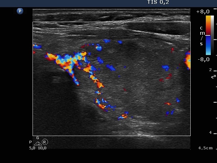
Middle part of the left lobe, horizontal scan, color Doppler mode. Signs of type 2 vascular pattern.

|
|
|
|
Middle part of the left lobe, horizontal scan, color Doppler mode. Signs of type 2 vascular pattern.