
|
|
100 consecutive patients with thyroid nodule - Case 18.
|

|


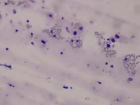

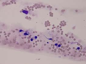
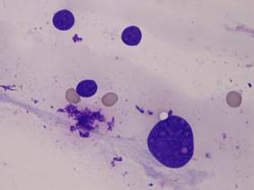
Clinical presentation: a 60-year-old woman was referred for aspiration cytology. She underwent a left lobetcomy for 7 seven years. 6 months before the present investigation a colon carcinoma was found.
Palpation: no abnormality.
Functional state: euthyroidism (TSH 2.01 mIU/L).
Ultrasonography. The right thyroid was moderately hypoechogenic and inhomogeneous. A lesion with a similar echopattern was found in the lateral part of the lobe. The mass presented halo sign. There was no parenchyma in the left thyroid bed.
Cytology resulted in benign lesion.
We performed wash-out thyrooglobulin examination which yielded 458 ug/L.
Suggestion: follow-up examination next time 3 years later.
Comment.
-
Thyrocytes presented pleomorphims. Nevertheless, there was no pathognomic atypia present, follicular cells exhibited neither inclusion nor groove nor oxyphilic changes. Atypia is a very common finding in thyroid smears. Simple hormonal influences not reflected in a blood test can cause such phenomenon. The rational question is in such cases is whether the atypia might correspond to any type of thyroid carcinoma. The arrangement of cells and the clinical history should be also taken into account. The lack of folliculus formation excluded a follicular-type tumor while the clinical presentation did an anaplastic carcinoma.
-
Considering the history, a metastasis had to be excluded, too. We did it with a wash-out thyroglobulin determination.


