
|
|
100 consecutive patients with thyroid nodule - Case 22.
|
|


Clinical data: a 39-year-old woman requested an evaluation because of a lump in the neck discovered by herself for several weeks.
Palpation: a very firm nodule in the right side of the isthmus.
Functional state: euthyroidism (TSH 1.86 mIU/L).
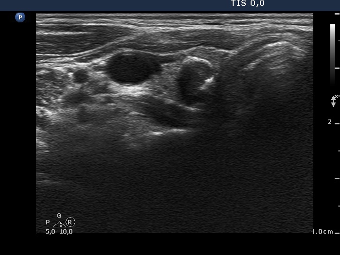
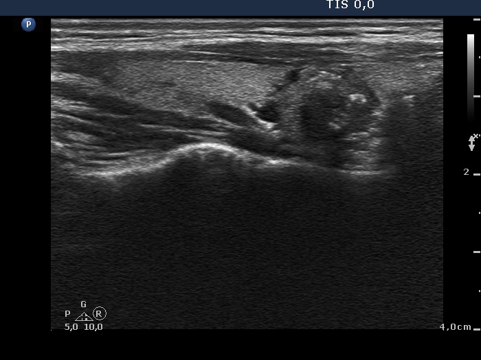
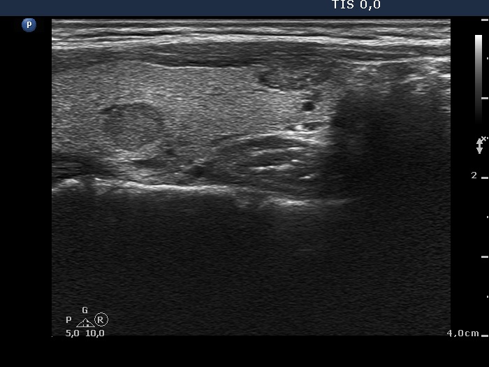
Ultrasonography. The thyroid was echonormal and contained multiple nodules including a hypoechogenic one in the lower pole of the right lobe presenting coarse calcifications. The nodule in the lower part of the left lobe was also noteworthy.
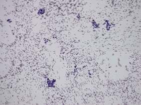
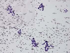
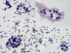
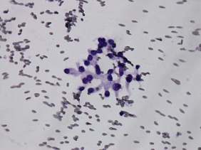
Cytology was performed form the nodule presenting microcalcifications and from that located in the lower pole of the left lobe. The latter resulted in benign colloid goiter while the cytology of the right nodule corresponded to a follicular tumor.
The common ultrasound-cytological diagnosis of the nodule in the right lobe was benign follicular proliferation.
Suggestion: ultrasound examination in two years.
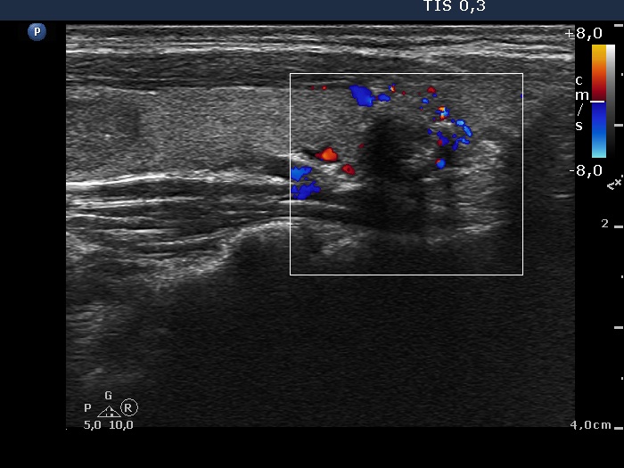
Comment. Although tht cytological pattern of the right lesion corresponds to a follicular tumor if we consider the ultrasound presentation than the risk of a follicular neoplasia is very low: the lesion exhibited neither halo sign nor perinodular bloodf flow. It means that there were not present any signs of a capsule. In such cases the risk of a follicular tumor is less than 5%.