
100 consecutive patients with thyroid nodule - Case 22. (ultrasonographic picture 7)
|
|

|
|
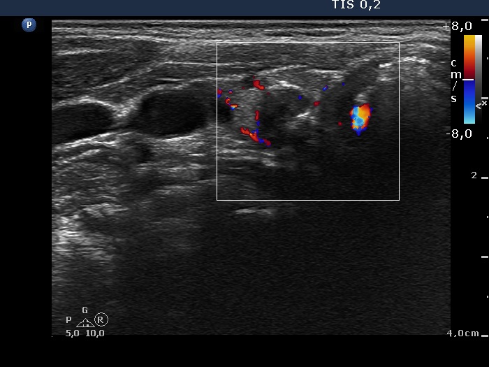
Lower pole of the right lobe, horizontal scan, color Doppler mode. A type 1 vascular pattern, i.e. neither perinodular nor intranodular blood flow can be detected.