
100 consecutive patients with thyroid nodule - Case 24. (ultrasonographic picture 8)
|
|

|
|
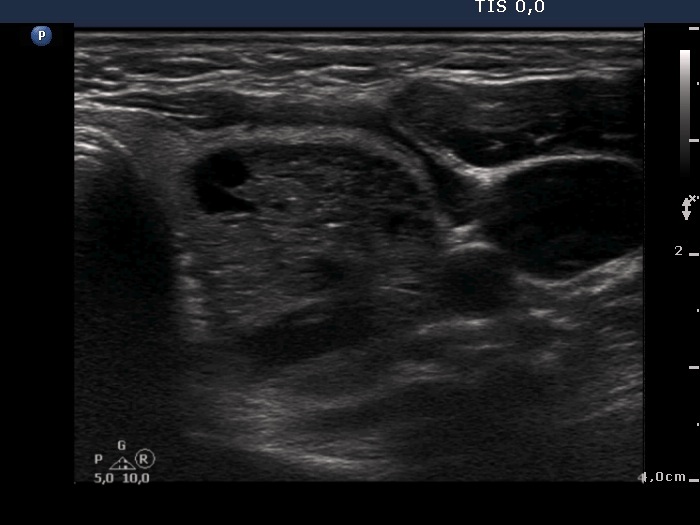
Left lobe, horizontal scan. Theupper part of the nodule contains a moderately hypoechogenic solid part presenting numerous hyperechogenic figures.