
|
|
100 consecutive patients with thyroid nodule - Case 29.
|
|


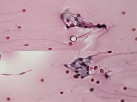
Clinical data: a 48-year-old woman was referred for evaluation of a nodular goiter discovered by chance on evaluation of arrhythmia.
Palpation: an elastic nodule in the left lobe.
Functional state: euthyroidism (TSH 0.76 mIU/L).
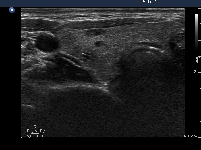
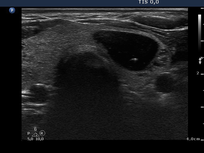
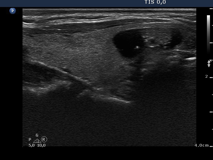
Ultrasonography. The thyroid was echonormal and contained several small cystic lesions in the right lobe and a relatively larger cystic nodule in the left lobe. The latter, peripheral-type cyst presented comet-tail artifacts and connective tissue in the moderately hypoechogenic solid part.
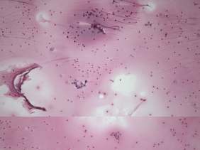
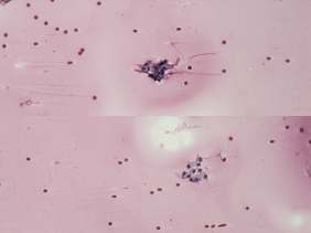
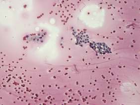
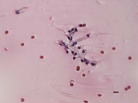
Cytology of the thyroid lesion resulted in benign colloid goiter.
Suggestion: follow-up examination in a year.
Comment. It is worth to analyze the hyperechogenic figures. The cystic area has various forms of colloid crystals while the appearance of the figures in the solid part might be misleading. There are both hyperechogenic lines and granules, therefore it should be avoided to interpret these as microcalcifications; these are presentation of connective tissue.