
100 consecutive patients with thyroid nodule - Case 35. (ultrasonographic picture 1)
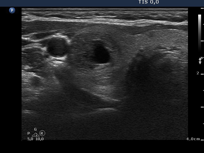 Right lobe, horizontal scan. There is a minimally-moderately hypoechogenic nodule presenting a central cystic degeneration. |
100 consecutive patients with thyroid nodule - Case 35. (ultrasonographic picture 1)
Right lobe, horizontal scan. There is a minimally-moderately hypoechogenic nodule presenting a central cystic degeneration. |

