
100 consecutive patients with thyroid nodule - Case 35. (ultrasonographic picture 6)
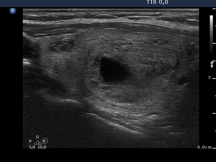 Left lobe, horizontal scan, color Doppler mode. The left lobe presents non-specific vascularization while the perinodular blood flow of the nodule in the right lobe can be seen well. |

