|
|||||||||
|
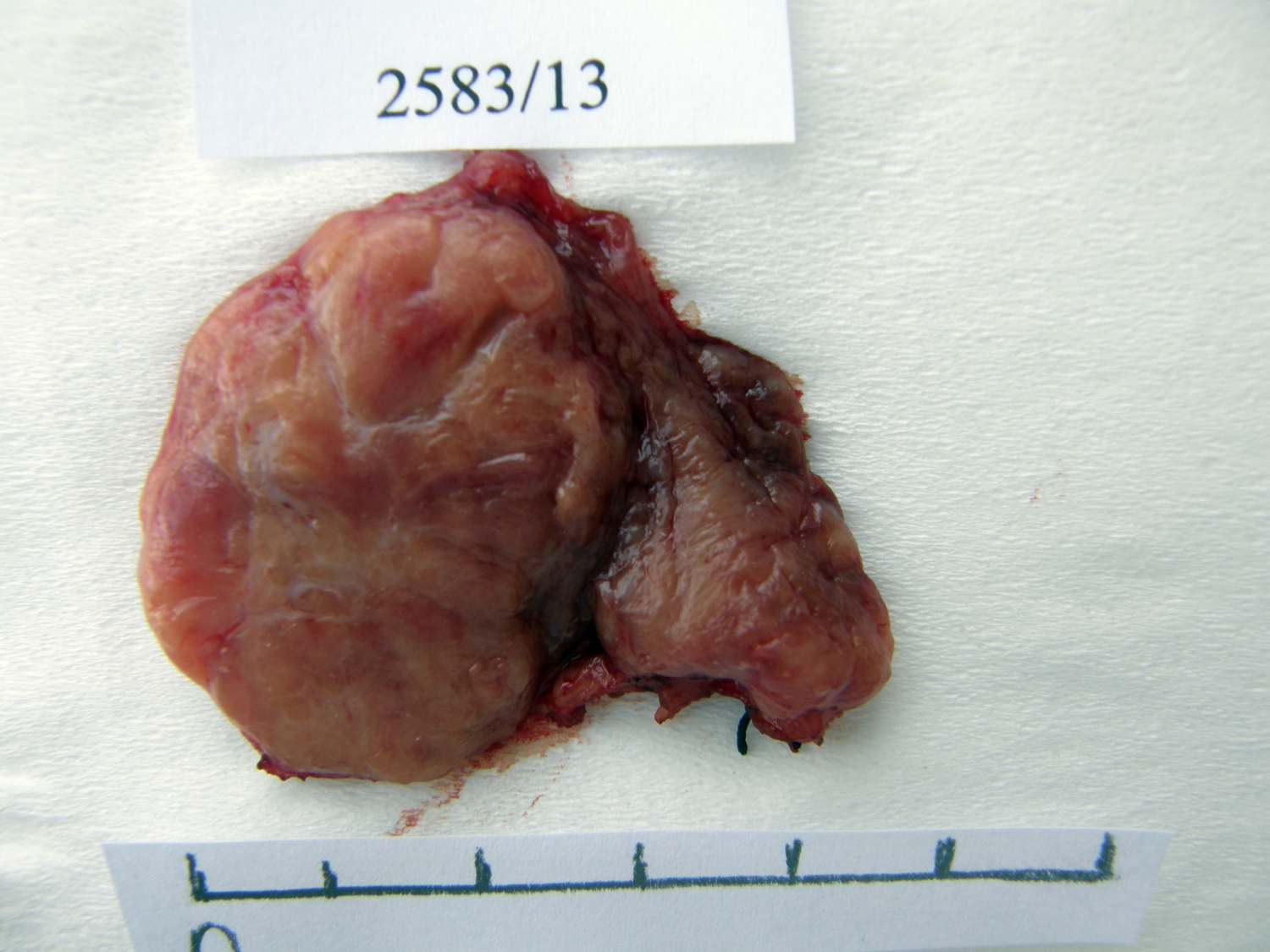
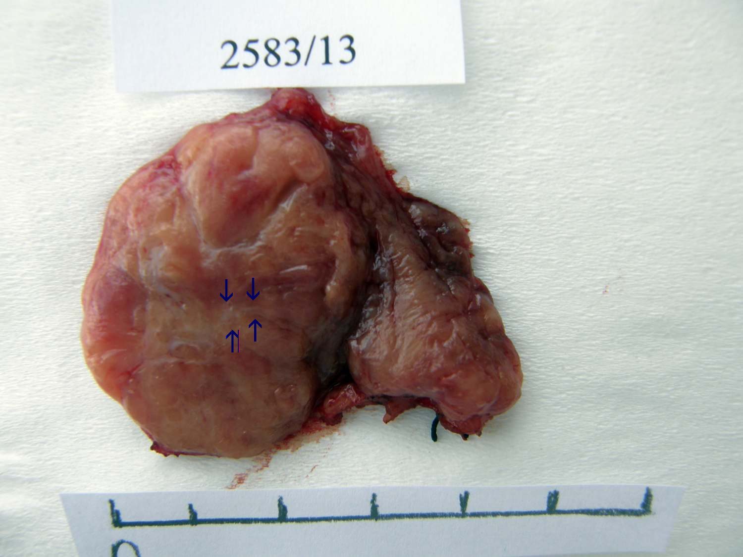
Macroscopic presentation of thyroid diseases. Table 15: An embryonal adenoma presenting Hashimoto's thyroiditis and a focus of papillary carcinoma.
|
|||||||||
|
|||||||||
|
|||||||||
|
Macroscopic presentation of thyroid diseases. Table 15: An embryonal adenoma presenting Hashimoto's thyroiditis and a focus of papillary carcinoma.
|
|||||||||
|
|||||||||