
|
|
Secondary thyroid carcinomas - Case 7.Metastasis of a small cell lung cancer
|
|

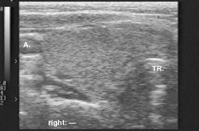


Clinical data: a 48-year-old woman who had been treated for Graves-Basedow's disease attended a follow-up examination. The patient had been examined 2 months earlier, when US was performed and revealed a nearly normal echo pattern without any discrete echo abnormality (the first US picture). She was then well. 2 weeks before the present examination she felt dyspnea, which progressed over the next 2 weeks.
Palpation: a moderately enlarged thyroid and an enlarged, hard cervical lymph node on the right were palpable.
Functional state: euthyroidism.
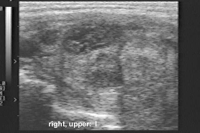
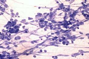
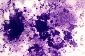
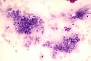
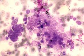
Ultrasonography: besides the enlarged cervical lymph node, US revealed a hypoechogenic inhomogeneous nodule in the right lobe of the thyroid (the second US picture).
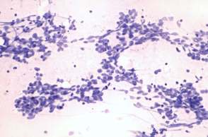
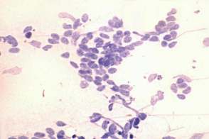
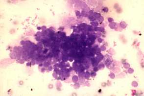
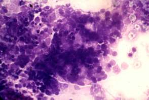
Cytology was performed. The FNAC result on both lesions (palpation-guided on the lymph node, and US-guided on the thyroid) was malignancy, i.e. a suspicion of small cell lung cancer, which was confirmed by immunocytochemistry; the tumor cells did not express LCA, but gave a positive reaction for neuron-specific enolase.
The patient was referred for a pulmonary evaluation. Bronchoscopy revealed a tumor in the lower lobe of the right lung.
Bronchoscopic brush cytology: gave the diagnosis of small cell lung cancer.
Comment: the possibility of a medullary carcinoma had to be considered on FNAC of the thyroid.