
|
|
Chronic lymphocytic thyroiditis - Case 5.
|
|

Clinical presentation: a 57-year-old woman was referred for aspiration cytology of a suspicious nodule. She visited her family physician with complaints suggesting hypothyroidism. Blood test and ultrasonography were performed. Hormonal examination disclosed hypothyroidism. The radiologist told the patient that she had a highly suspicious nodule which was with great probability a malignant one.
Palpation: both thyroids were firm on palpation.
Functional state: hypothyroidism with TSH 10.9 mIU/L, FT4 8.94 pM/L.
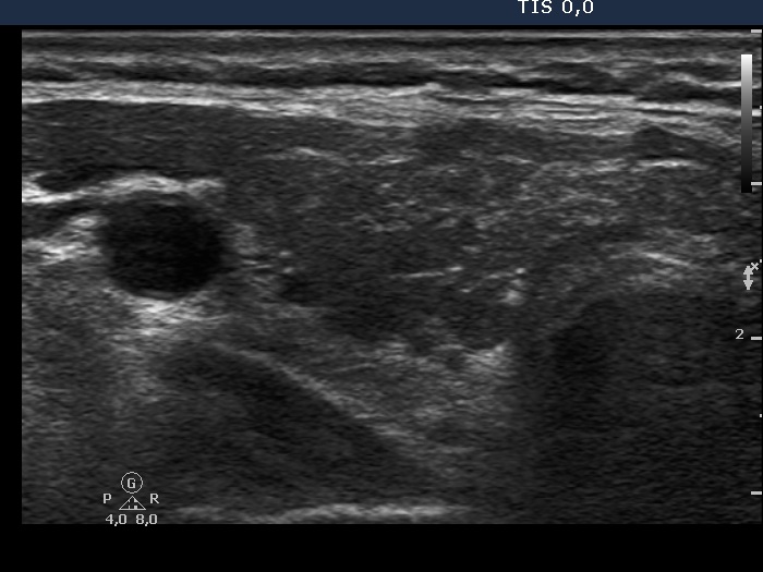
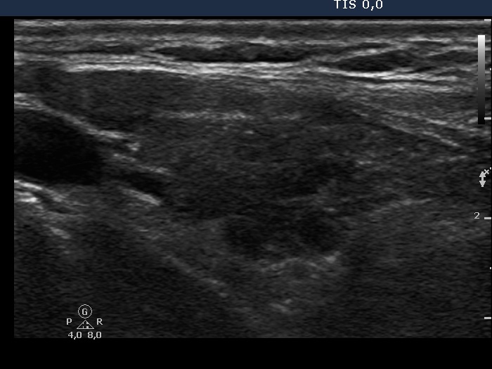
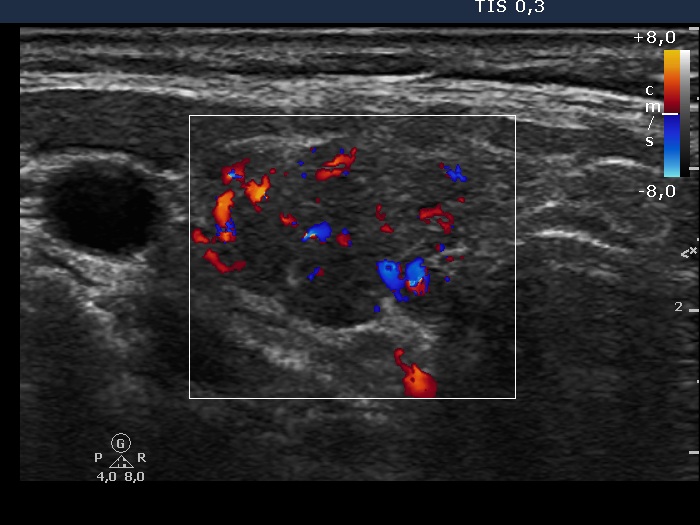
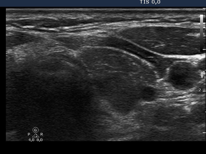
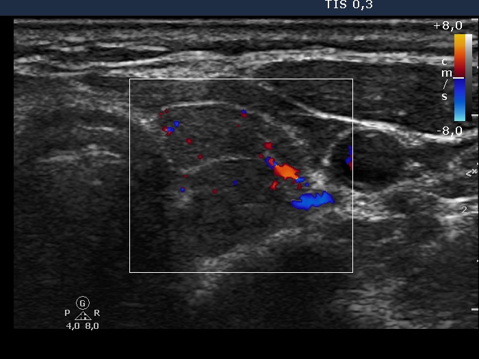
Ultrasonography: both thyroids were hypoechogenic. The echogenicity index was 90% in the right, and 100% in the left thyroid. The hypoechogenic area was surrounded with a small echonormal rim in the right lobe. This pattern, which is very specific for lymphocytic thyroiditis, was misinterpreted as a large nodule by the former examiner. (See details in comment to the first ultrasound picture.) There were multiple more hypoechogenic areas within the thyroid, none of them corresponded to nodule.
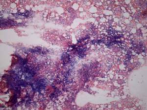
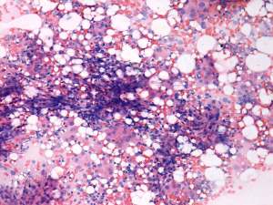
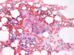
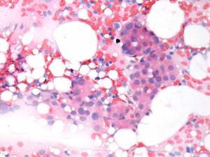
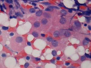
FNAC: disclosed Hashimoto's thyroiditis.
The patient was very anxious because of the first ultrasound report and she wanted to be operated. We could not persuade the patient not to undergo surgery.
Histopathology: disclosed Hashimoto's thyroiditis.