
The borders of the nodule - case 349 (cytologic picture 1)
|
|

|
 |
|
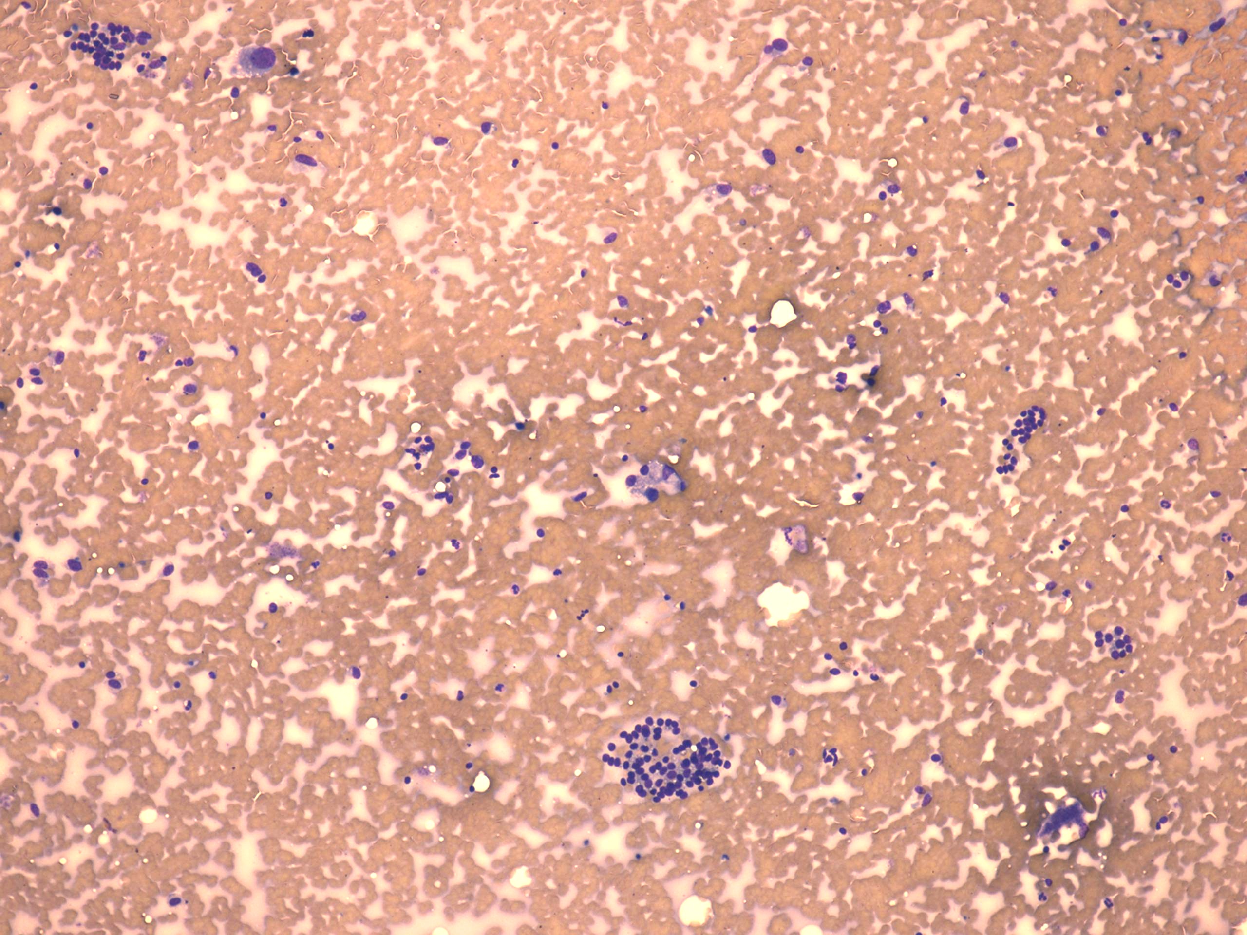
Wright-Giemsa-staining, 100x. Two cell populations are demonstrated: there is a group of regular follicular cells in the lower part of the image while a cluster of atypical cells and an enlarged, isolated cell in the left-upper part of the image.