
|
|
The borders of the nodule - case 808
|
|


Clinical presentation: A 36-year-old woman was referred for evaluation of an elevated TSH (5.08 mIU/L) detected on routine blood test 2 months ago. She had no complaints. The height of the patient was 161 cm while the weight was 42 kg.
Palpation: a moderately firm nodule in the right lobe.
Functional state: euthyroidism with TSH 2.18 mIU/L, FT4 14.3 pM/l, aTPO 0 U/mL.
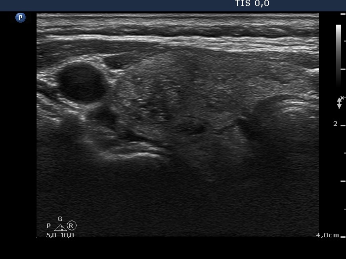
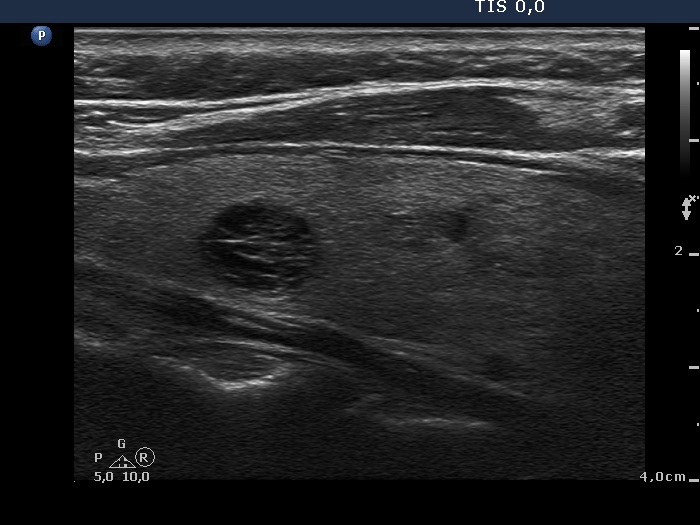
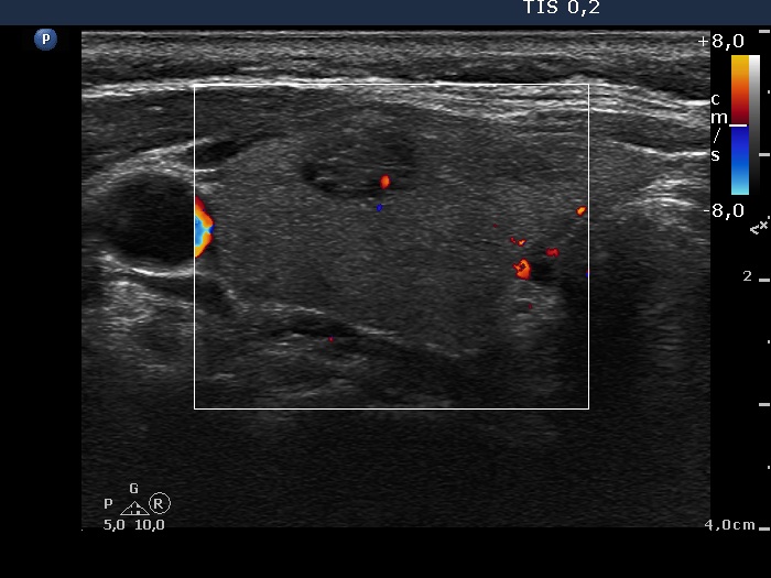
Ultrasonography. The thyroid was echonormal. There were several small nodules in both lobes which presented cystic degeneration. There was a cystic nodule in the isthmus. The solid part of the lesion contained various hyperechogenic granules and lines.
Cytology resulted in benign cystic-colloid goiter.
Comments.
-
The cause for elevated TSH remains unclear. There were no signs of an underlying thyroiditis, neither the ultrasound nor the aTPO nor the cytology suggested autoimmune thyroiditis. On the other hand, the physique of a patient has a well-known impact on the normal value of TSH, thinner the build higher the upper level of TSH.
-
At first sight the two bright hyperechogenic granules presented in the transverse scan of the right lobe seems to be difficult to categorize. However, the less bright granules and the synchronous presence of hyperechogenic lines are signs of connective tissue. Therefore, the bright granules belong very likely even to this subgroup.
- Regarding nodule borders. The nodule in the upper part of the right lobe seems to have blurred borders. However, this blur is caused only by the similar echogenicity of the nodule and the extranodular part.