
The borders of the nodule - case conp 027 (cytologic picture 10)
|
|

|
 |
|
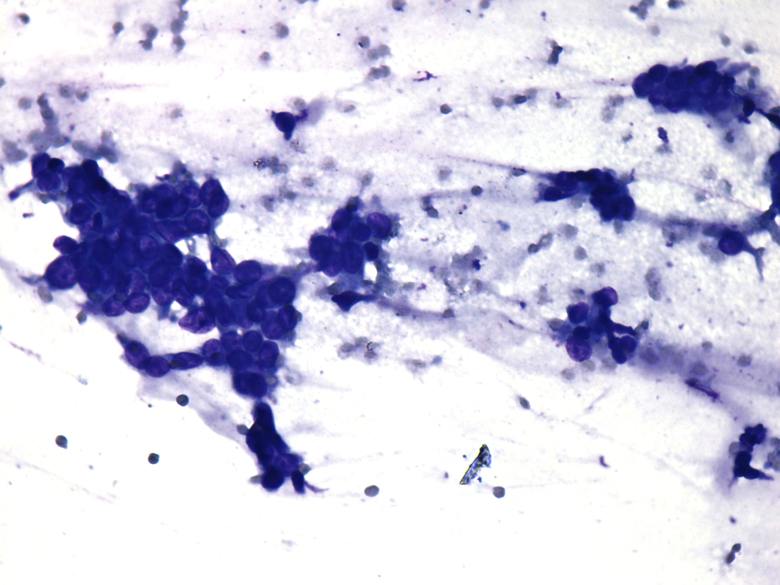
Wright-Giemsa staining, 400x. There is an irregular cluster, which might correspond to part of a papillary fragment. The nucleus in the left central part of the image presents a groove.