
|
|
The composition of the nodulen - case 399
|
|


Clinical presentation: A 47-yr-old woman was referred for aspiration cytology. She noticed a lump in her right thyroid lobe four month ago. Three mL cystic fluid was aspirated in another hospital. FNA resulted in benign cystic lesion. The cyst has relapsed within a few weeks. Among others, calcitonin test was performed and yielded twice an elevated levels (21.2 and 28.9 pg/mL - normal value below 11 pg/mL).
Palpation: an elastic nodule in the right lobe.
Laboratory tests: TSH 2.69 mIU/L, aTPO 0.5 U/mL, calcitonin 28.9 pg/mL.
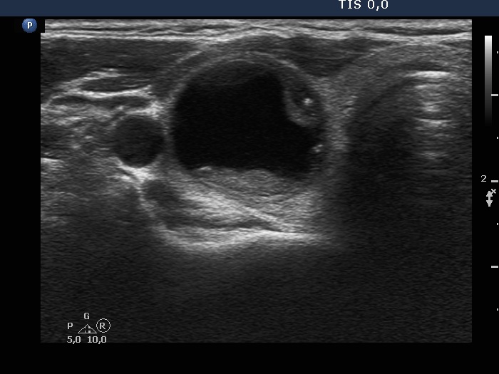
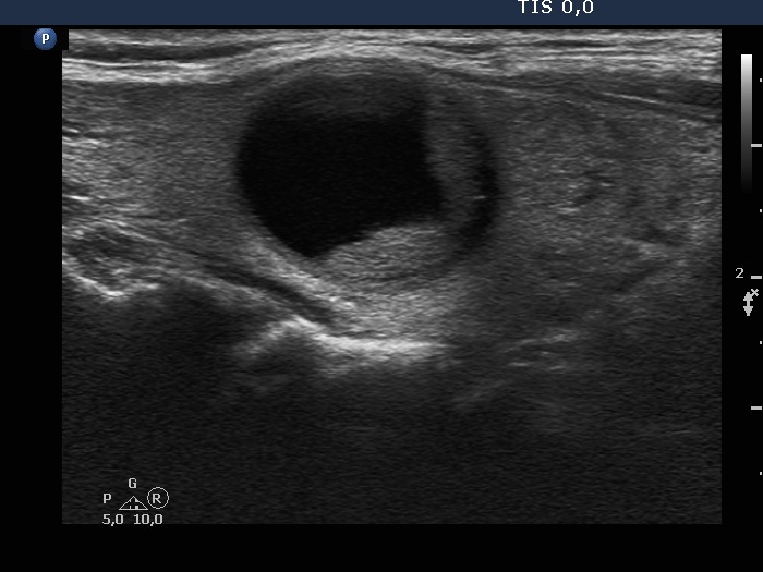
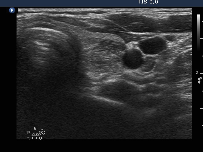
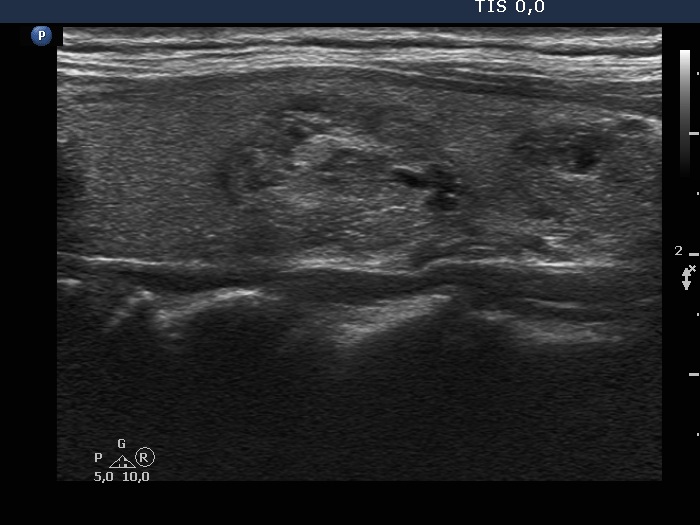
Ultrasonography. The thyroid was echonormal and had multiple moderately hypoechoic and hyperechoic nodules which showed various degrees of cystic degeneration. The largest nodule in the right lobe was dominantly cystic and presented with minimally hypoechoic solid part which included hyperechoic figures. The largest nodule in the left lobe had irregular margins.
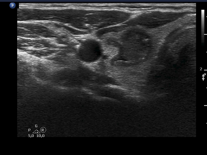
2.5 mL cystic fluid was aspirated from the right nodule. Thereafter, the borders became irregular, lobulated. Aspiration cytology of the solid part after the removal of cystic fluid an that of the nodule having irregular borders in the left lobe resulted in bening cystic degeneration.
The endocrinologist of the patient suggested total thyroidectomy.
Histopathology resulted in benign hyperplastic nodules.
I am aware of three more calcitonin determinations performed after the surgery. These tests resulted in 17.2 pg/ml, 2.4 pg/mL and 20.9 pg/mL.
Comment.
-
There were many deceptive circumstances in this patient.
-
The repeatedly elevated calcitonin levels. Considering the postoperative results, this elevated levels area very likely of non-pathological cause.
-
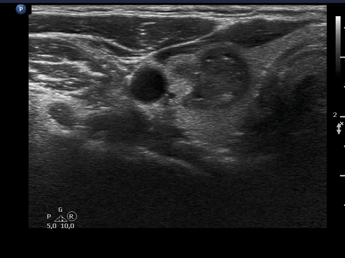
The right nodule presented with a patchy echonormal area having punctate echogenic foci, a pattern resembling amyloid deposit.
-
The left nodule had irregular borders.
-
-
The nodule' borders became irregular after aspiration of cystic fluid. This is a phenomenon which should not be regarded as a pathological finding.