
|
|
The composition of the nodule - case 723
|
|


First examination (first row of images):
Clinical presentation: A 31-year-old woman was referred for evaluation of a nodule discovered by herself.
Palpation: a not firm nodule in the isthmus.
Hormonal evaluation: euthyroidism with TSH 0.75 mIU/L.
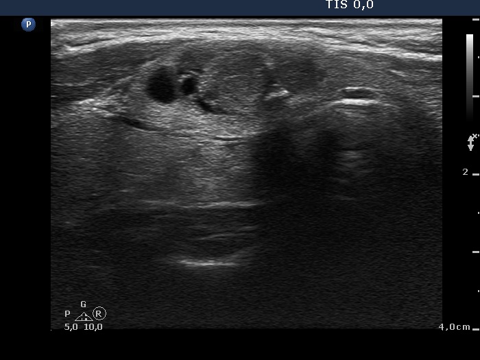
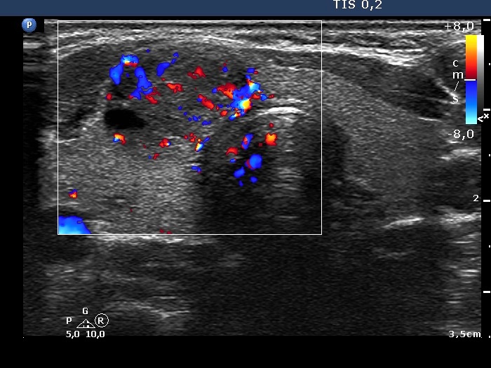
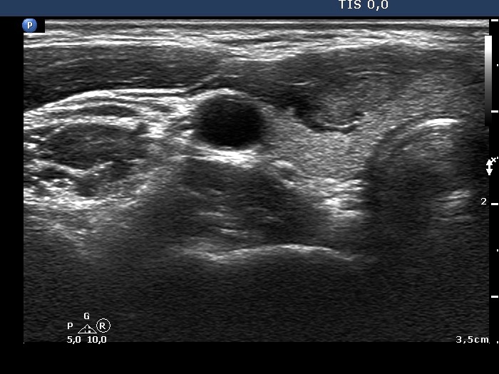
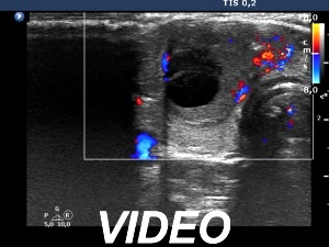
Ultrasonography. The thyroid was echonormal. There was a small, moderately hypoechogenic lesion in the lower part of the left lobe while a larger nodular area was located in the isthmus. The latter was composed of hypoechogenic and echonormal parts and presented cystic degeneration, as well.
Aspiration cytology of the isthmic nodule disclosed a follicular lesion with signs of hyperthyroidism.
We indicated scintigraphy which revealed an autonomously functioning nodule according to the lesion in the isthmus. We suggested follow-up, yearly TSH determination.
One year after the first examination (second row of images):
Clinical presentation. The patient had no complaints.
Palpation: unchanged.
Hormonal evaluation: TSH 0.91 mIU/L.
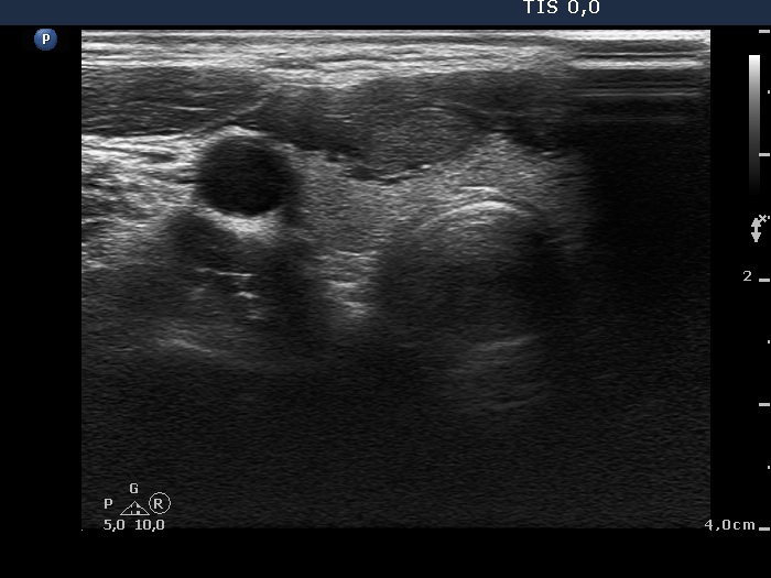
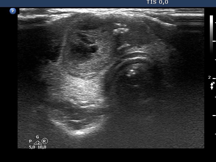
Ultrasonography. The cystic areas of the nodule in the isthmus have increased. The lesion presented more bright echogenic lines and granules which corresponded to figures caused by posterior back wall enhancement.
Four years after the first examination (third row of images):
Clinical presentation. The patient had no complaints.
Palpation: unchanged.
Hormonal evaluation: TSH 0.72 mIU/L.
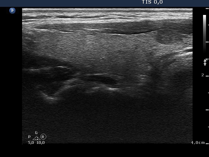
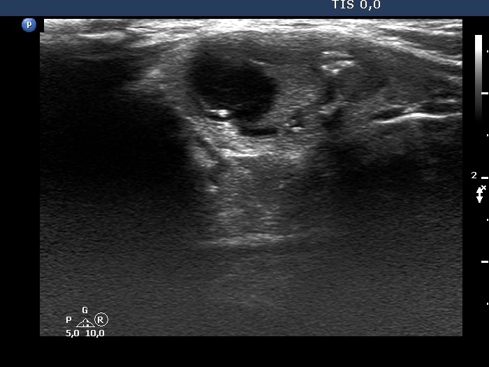
Ultrasonography. The lesion increased by 35% larger in volume compared with the first examination, primarily because of the increase of the cystic content.
Suggestion: TSH in a year, ultrasound in 3 years.