
Intranodular hyperechogenic figures - case 41
Follow-up examination 2 years later (ultrasonographic picture 2)
|
|

|
|
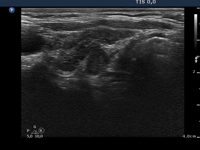
Right lobe, longitudinal scan. There are several discrete lesions divided by fibrous septa.