Intranodular hyperechogenic figures - case 41
Follow-up examination 2 years later (ultrasonographic picture 3)
|
|

|
|
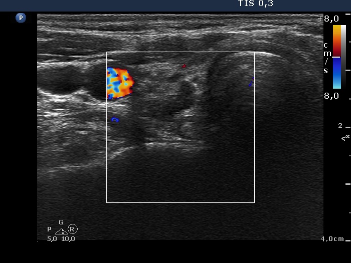
Right lobe, transverse scan, color Doppler mode. The vascularization is decreased.

2022-23 Advanced
Papillon Course
Echogenic figures
|
|
|
|
Right lobe, transverse scan, color Doppler mode. The vascularization is decreased.