Intranodular hyperechogenic figures - case 694 (ultrasonographic picture 15)
|
|

|
|
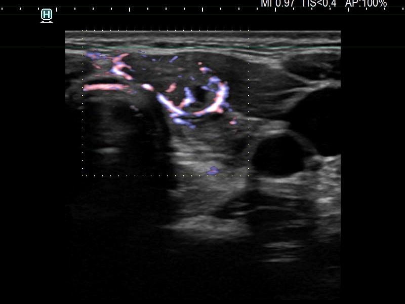
Left lobe, transverse scan, fine flow method. The lesion presents a combined perinodular and intranodular blood flow.

2022-23 Advanced
Papillon Course
Echogenic figures
|
|
|
|
Left lobe, transverse scan, fine flow method. The lesion presents a combined perinodular and intranodular blood flow.