
Lymphocytic thyroiditis - case 1442
Follow-up investigation 5 years after the first visit (ultrasonographic picture 1)

|
|
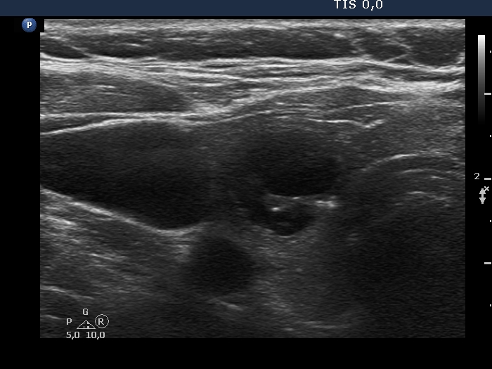
Right lobe, transverse scan. The central cystic content has gathered in the lesion. The previously pale granules and lines became bright. These are located dorsal to tiny or larger cystic areas, therefore they correspond to back wall cystic figures.