
|
|
Lymphocytic thyroiditis - case 1442
|
|

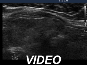
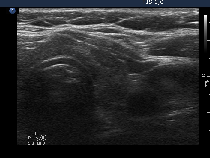
Examination in 2013 (first row of images):
Clinical data. A 50-year-old woman was referred for evaluation of a nodular goiter. The patient has been treated for hypothyroidism for 16 years.
Palpation: Both lobes were a bit firm, no nodule was palpable.
Laboratory test: TSH 4.01 mIU/L on daily 100 microgram levothyroxine.
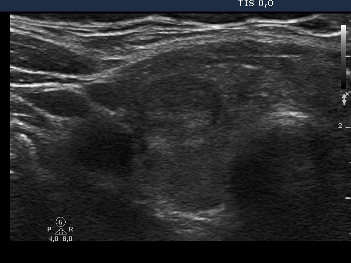
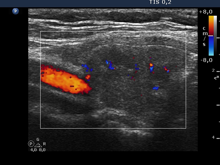
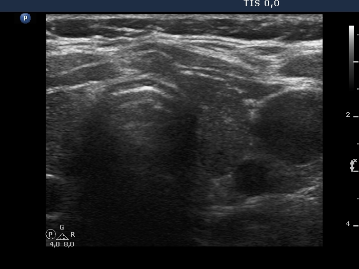
Ultrasonography. The thyroid was minimally/moderately hypoechoic and had several discrete minimally/moderately hypoechoic lesions.
Cytology was performed from the lesion in the central part of the right lobe and resulted in Hashimoto's thyroiditis.
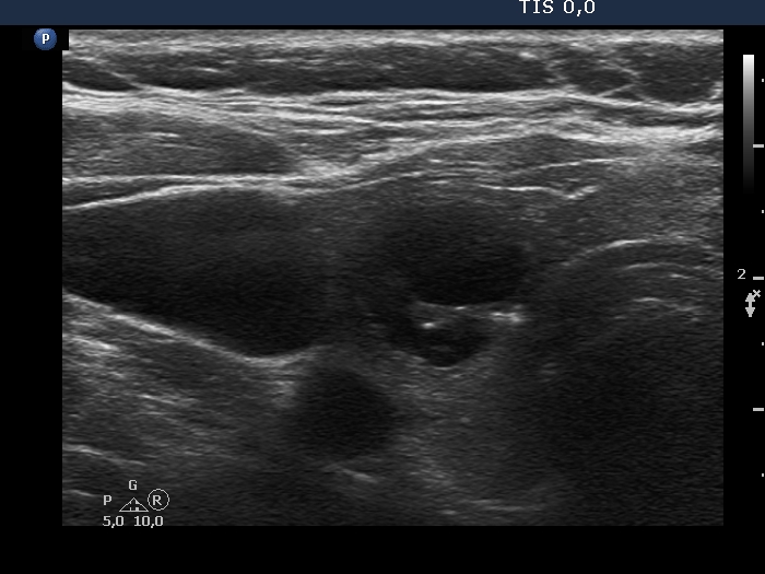
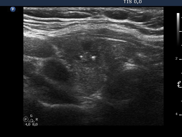
Examination in 2018 (second row of images):
Clinical data. The patient came to a routine follow-up. She had no complaints.
Palpation: unchanged.
Laboratory test: TSH 0.36 mIU/L on daily 125 microgram levothyroxine.
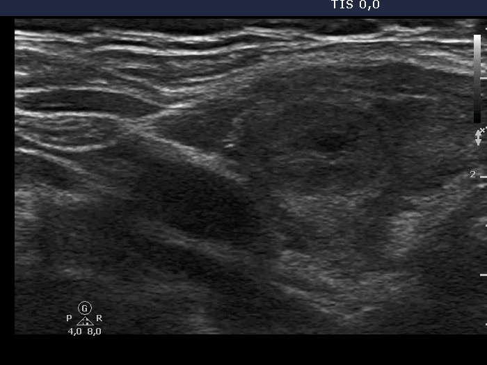
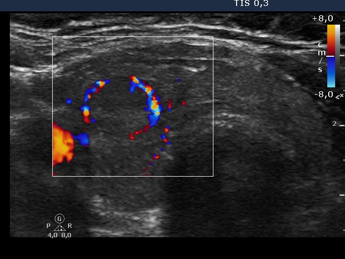
Ultrasonography. Except for the lesion which was previosuly cytologically investigated, the pattern remained unchanged. The lesion in question became cystic and was composed of two chambers. In the tissue part separating the two chambers, hyperechoic granules have appeared. These were mostly related to ventral cystic areas, therefore, they should be regarded as back wall figures.
We recommended that she takes the replacement therapy at the same dose and has a TSH scan after half a year and an ultrasound scan after two years.
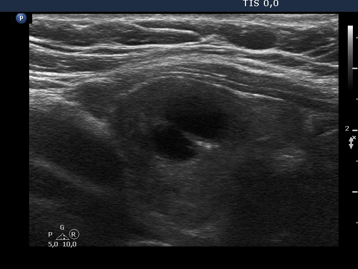
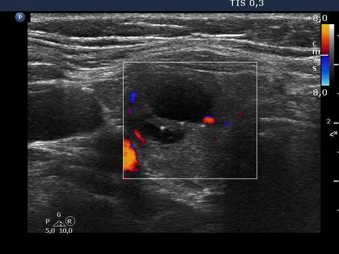
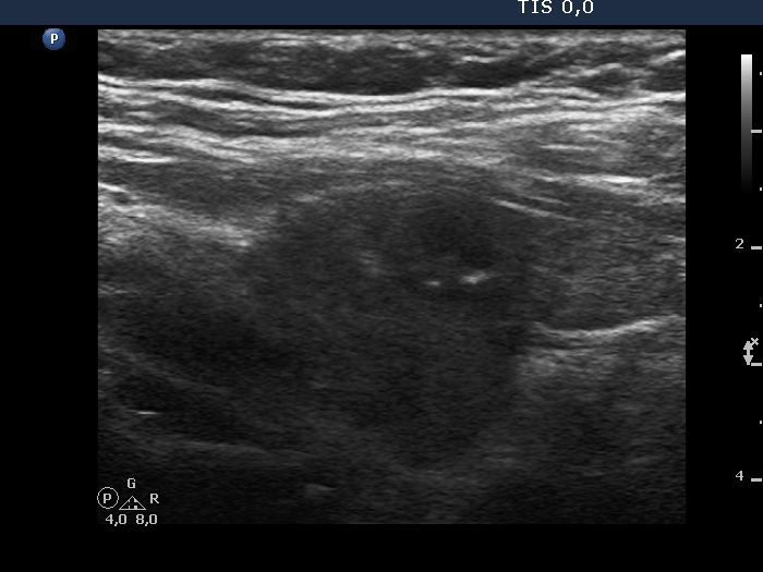
Examination in 2020 (third row of images):
Comment. This story draws attention to how important it may be to be aware of the results of a previous study. By result I do not mean the findings, but the archived recordings.Clinical data: The patient came to a routine follow-up. She had no complaints.
Palpation: Both lobes were a bit firm on palpation. There was a not firm nodule in the right lobe.
Laboratory test: TSH 3.38 mIU/L on daily 125 microgram levothyroxine.
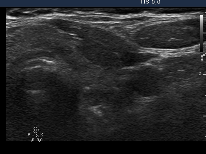
Ultrasonography. The previously cystic lesion has spontaneously decreased in size, the cystic fluid has disappeared. However, the hyperechogenic foci were still visible. If we had only seen this current study, we would have had to regard these echogenic granules microcalcifications.
We recommended that she takes the replacement therapy at the same dose and has a TSH scan after a year and an ultrasound scan after three years.