
Lymphocytic thyroiditis - case 1442
Follow-up investigation 5 years after the first visit (ultrasonographic picture 3)
|
|

|
|
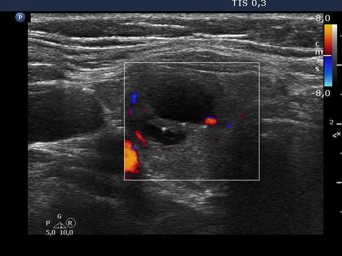
Right lobe, transverse scan, color Doppler method. The lesion shows signs of perinodular vascularization.