
Lymphocytic thyroiditis - case 1442
Follow-up examination 7 years after the first one (ultrasonographic picture 1)

|
|
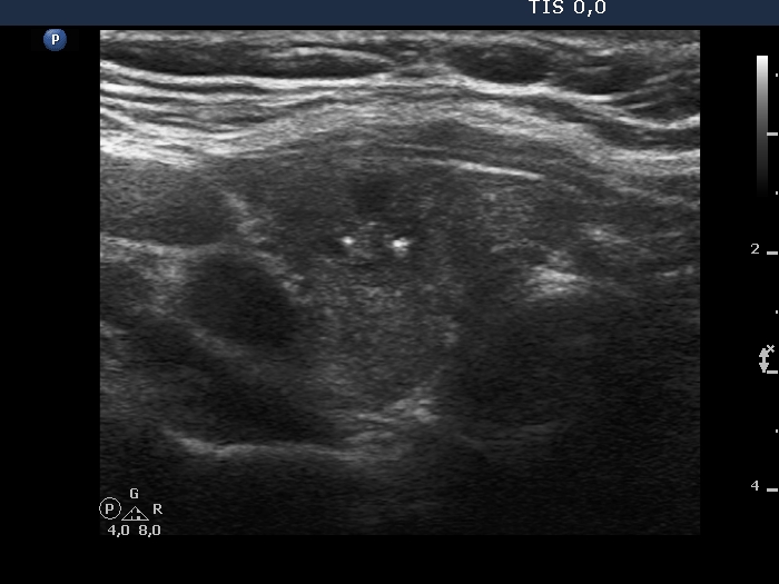
Right lobe, transverse view. The cystic fluid has almost completely disappeared but not the echogenic figures. Although they are dorsal to tiny cystic areas, solely on this presentation, microcalcification cannot be excluded but is even probable. Fortunately, thanks to the archived images and videos, we could avoid a false interpretation.