
|
|
Case 1738
|
|

Clinical data: A 25-year-old woman was referred for evaluation of a suspicious nodule which was detected on routine ultrasound examination. The patient has undergone on total thyroidectomy and radioiodine therapy for a papillary cancer for 6 years. The tumor was a T2 cancer. Thyroglobulin levels were in the range of 0.2 to 0.8 ng/dL throughout the postoperative period without any tendency.
Palpation: no abnormality.
Laboratory tests: TSH 0.09 mIU/L, FT4 20.5 pM/L, thyroglobulin 0.6 ng/mL, anti-hTg 15 U/mL on daily 112.5 microgram levothyroxine.
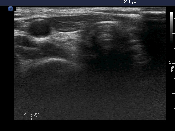
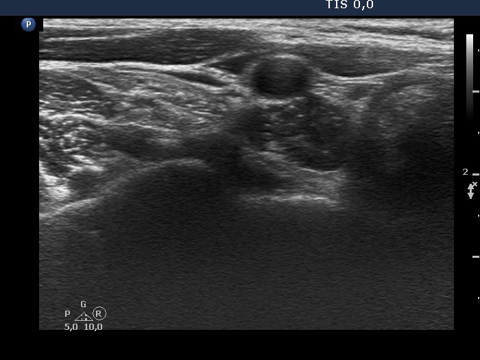
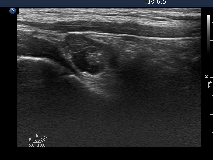
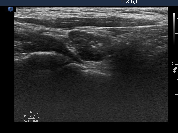
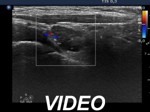
Ultrasound. Connective tissue replaced the thyroid parenchyma in both thyroid beds. There was a hypoechoic mass in the lower part of the right thyroid bed dorsal to the carotid artery. The lesion was a mixed, dominantly solid nodule and had microcalcifications.
Aspiration cytology from the hyperechogenic lesion resulted in papillary cancer. Wash-out thyroglobulin was 6905 ng/mL
Suggestion: On surgical exploration 11 lymph nodes were removed from the right side of the neck. Two of them were metastatic.
Comment. This case study underlines the importance of minimal thyroglobulin levels.