
Case 1738 (ultrasonographic picture 4)
|
|

|
|
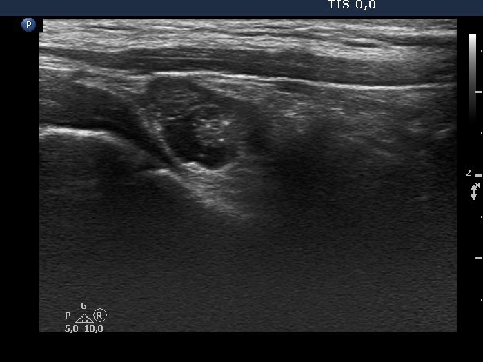
Right lobe, longitudinal view. This image proves that the nodule has cystic areas. It is worth comparing the presentations of the echogenic figures in the upper (left in the image) and in the lower (right in the image) solid parts. These correspond to back wall figures in the former while microcalcifications in the latter.