
Case 2007 (ultrasonographic picture 5)
|
|

|
|
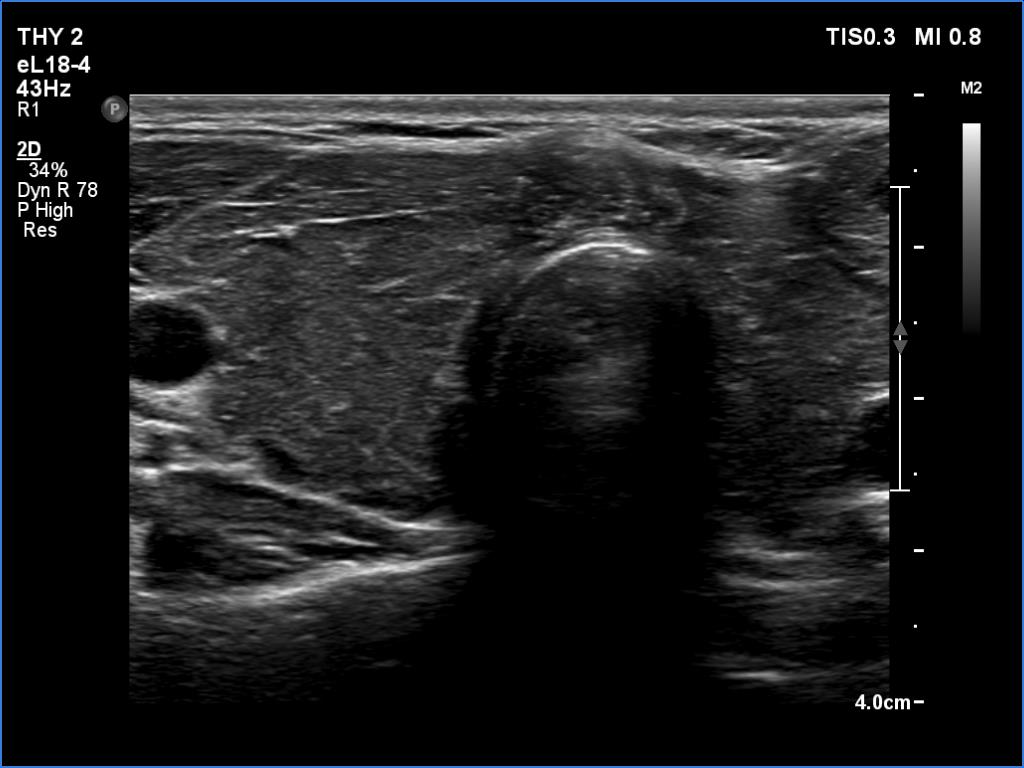
Isthmus, transverse scan. There is a more hypoechoic lesion in the isthmus. The punctate echogenic granules within the lesion are brighter than those in the extranodular tissue.