
Case 856
Follow-up investigation two years later (ultrasonographic picture 5)
|
|

|
|
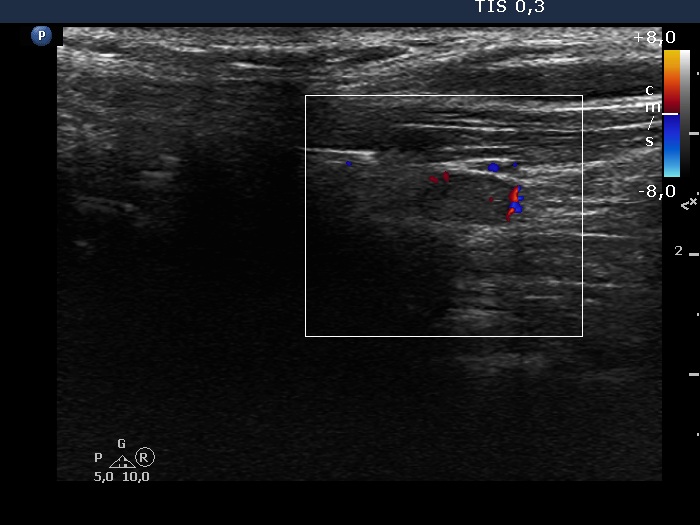
Left lobe, longitudinal scan, color Doppler mode. Although the vescularity is scanty, the demonstration of even a minimal vascularization also proves that the lesion is not a muscle fiber.