Case 856
Follow-up investigation two years later (ultrasonographic picture 6)
|
|

|
|
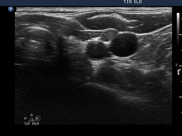
Left lobe and lateral to the left lobe, transverse scan. There is a hypoechoic mas ventral and between the vessels.

2022-23 Advanced
Papillon Course
Lesion vs. nodule
|
|
|
|
Left lobe and lateral to the left lobe, transverse scan. There is a hypoechoic mas ventral and between the vessels.