
Case 856
Follow-up investigation two years later (ultrasonographic picture 9)
|
|

|
|
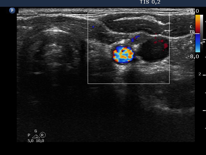
Left lobe and lateral to the left lobe, transverse scan, color Doppler mode. The scanty vascularity proves that the mass is not a muscle fiber.