
100 consecutive cases of papillary cancer - case 076 (ultrasonographic picture 14)
|
|

|
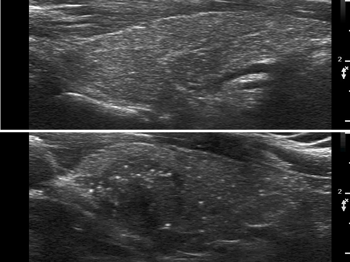
Comparison of the right lobe (upper) presenting fibrotic changes with the left lobe (lower) showing numerous microcalcifications. Note the difference between the size of the hyperechoic granules caused by fibrosis and microcalcifications.