
|
|
100 consecutive cases of papillary cancer - case 087
|
|


Clinical presentation: A 15-year-old girl came to a follow-up examination. We met her one year ago when her parents requested a second opinion. (On screening in the school, an enlarged thyroid was found. Thereafter, a benign nodule was diagnosed in another hospital.) We suggested ultrasound in a year.
Palpation: The right lobe had a firm nodule.
Result of blood test: TSH 2.54 mIU/L, aTPO 1.3 U/mL
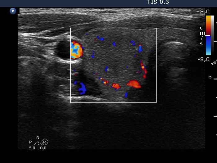
Ultrasonography. The thyroid was echonormal. There were two moderately hypoechoic nodules in the right lobe. They presented microcalcifications, the larger one ha irregular, lobulated margins. The nodule has grown significantly since the previous visit, from 8x8x15 mm to 14x12x20 mm, width, depth and length, respectively.
Cytology resulted in suspicion of papillary cancer.
Histopathology disclosed papillary carcinoma.