
|
|
100 consecutive cases of papillary cancer - case 088
|
|


Clinical presentation: A 46-year-old woman was referred for evaluation of a thyroid enlargement. The patient has been treated by levothyroxine for more than ten years. Six month prior to the present examination a hysterectomy was performed, histology resulted in myoma. The enlargement of the thyroid was discovered at that time.
Palpation: a firm nodule in the right lobe.
Laboratory test: TSH 0.04 mIU/L, FT4 24.7 pM/L on daily 175 microgram levothyroxine.
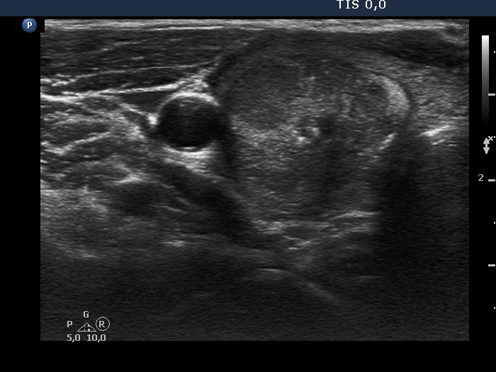
Ultrasonography. The thyroid was hypoechoic and presented with several discrete areas. A hypoechoic nodule occupied great part of the right lobe. The nodule had echonormal fields, microcalcifications and halo and presented perinodular blood flow.
Cytology resulted in papillary cancer.
Histopathology disclosed an encapsulated papillary cancer in the right lobe while multiple, non-encapsulated foci of papillary cancer in the left lobe. Hashimoto's thyroiditis was found in the extranodular part. The tumor in the right lobe infiltrated the thyroid' capsule in an area of 10 mm.