
|
|
100 consecutive cases of papillary cancer - case 089
|
|

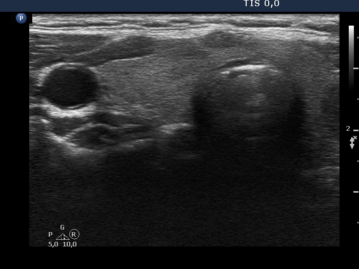


First examination (first row of images):
Clinical presentation: A 39-year-old woman was referred for evaluation of a nodule detected on ultrasound screening. Cytology resulted in benign lesion. The aTPO level proved to be elevated.
Palpation: a firm lesion in the left lobe.
Functional state: euthyroidism (TSH 2.65 mIU/L, anti-TPO > 1300 U/mL).
Ultrasonography. The thyroid was echonormal and has several small, moderately hypoechoic areas corresponding to thyroiditis. There was a hypoechoic nodule in the left lobe. The lesion had microcalcifications, presented with irregular, lobulated margins and showed a combined perinodular and intranodular vascularity.
Considering the ultrasound presentation, we performed cytology which resulted in non-diagnostic result.
Suggestion: ultrasound in a year.
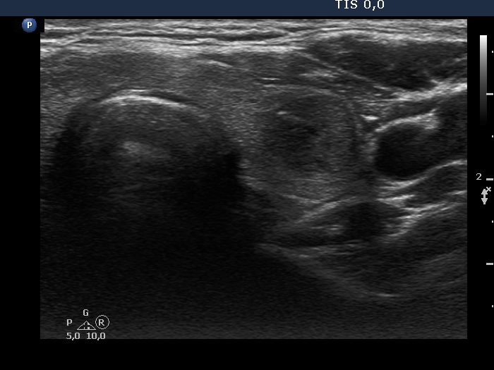
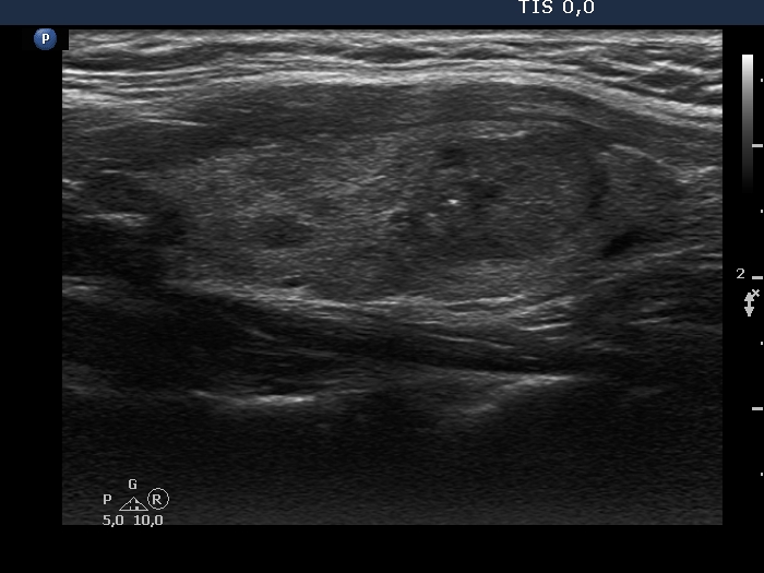
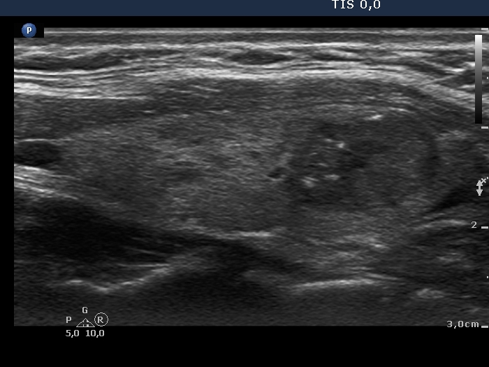
Second examination (second row of images):
Clinical presentation: The patient had no complaints.
Palpation: unchanged.
Functional state: euthyroidism (TSH 1.09 mIU/L).
Ultrasonography. The pattern remained unchanged.
Cytology resulted in Hashimoto's thyroiditis and suspicion of papillary cancer.
Total thyroidectomy was performed. Histopathology disclosed a T1b stage, encapsulated papillary cancer and Hashimoto's thyroiditis.
Comment. As a rule, we did not expect a malignant transformation of a benign nodule. The issue is the failure of FNA. In a nodule presenting suspicious ultrasound characteristics, repeat FNA is suggested even if the original cytological report was reassuring.