
100 consecutive cases of papillary cancer - case 100 (ultrasonographic picture 6)
|
|

|
|
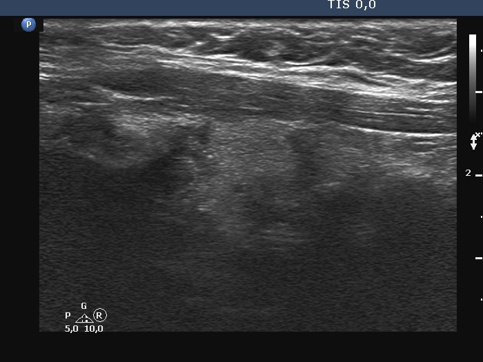
Left lobe, longitudinal scan. There at least three, relatively large discrete hypoechoic lesions. The middle one is suspicious due to the presence of microcalcifications and irregular borders.