
|
|
Introduction - case 1185
|
|


Clinical presentation: A 39-yr-old woman requested evaluation of a 'lump in the throat' feeling.
Palpation: no abnormality.
Laboratory test: TSH 0.85 mIU/L.
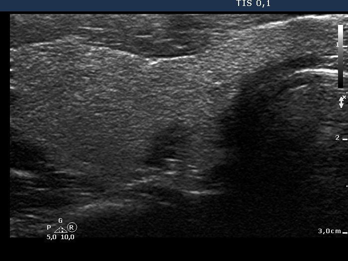
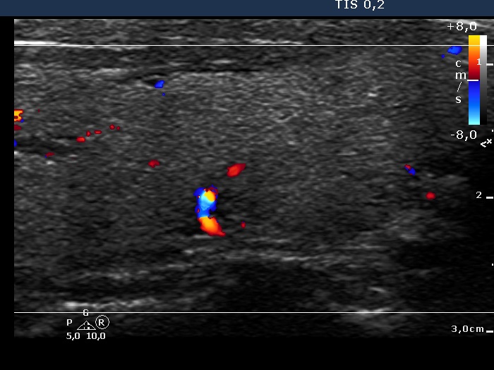
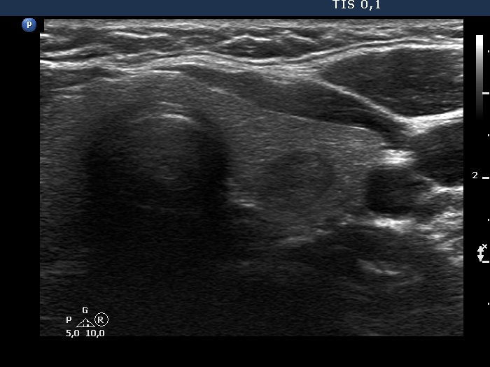
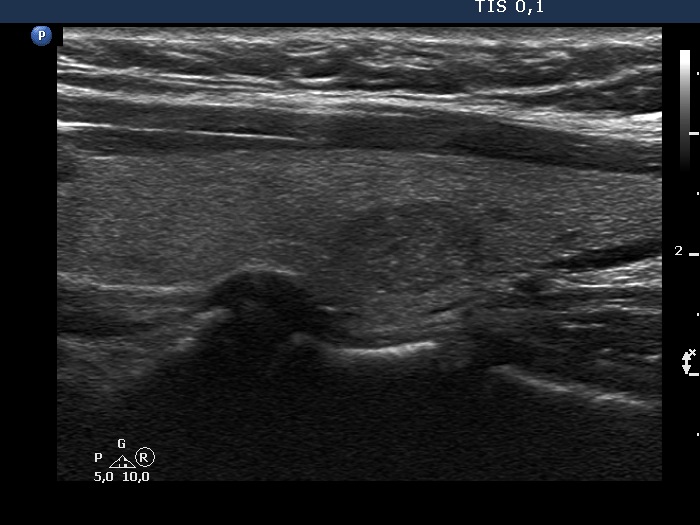
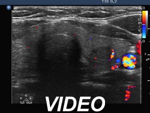
Ultrasonography. The thyroid was echonormal. There was a discrete, deeply hypoechoic area in the dorsal part of the right lobe. Although the dimensions were only 4x7x5 mm, width, depth, length, respectively, the lesion would correspond to a TIRADS 5 lesion because it was very hypoechoic and presented with irregular shape. However, color Doppler mode proved that this was indeed a vessel and not a solid thyroid tissue. The left lobe had a minimally hypoechoic lesion which largest diameter was 9 mm.
Suggestion. Ultrasound in 3 to 5 years due to the lesion in the left lobe.
Comments. Although some vascular patterns significantly increase the risk of malignancy, none of the TIRADS include vascularity among suspicious characteristics. The reason for this is the lack of standardization and the poor sensitivity and specificity. On the other hand, we must investigate the thyroid with color Doppler mode because we never know when to gain significant advantage of the technique. This case study is one of the many examples.