
Introduction - case 1326 (ultrasonographic picture 3)
|
|

|
|
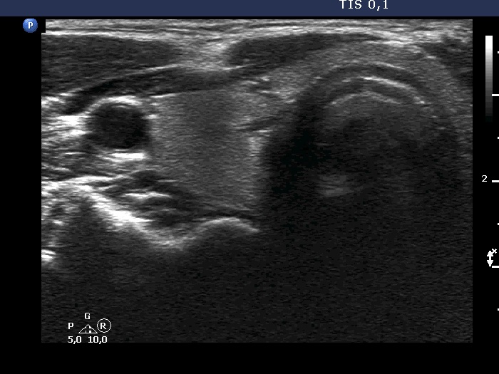
Right lobe, transverse view. In this section, the area mimics a moderately hypoechoic nodule having blurred margins. Note the thick connective tissue fragment within the strap muscle just ventral to the area.